New Imaging System Combines Ultrasound and Photoacoustic Tomography
New Imaging System Combines Ultrasound and Photoacoustic Tomography
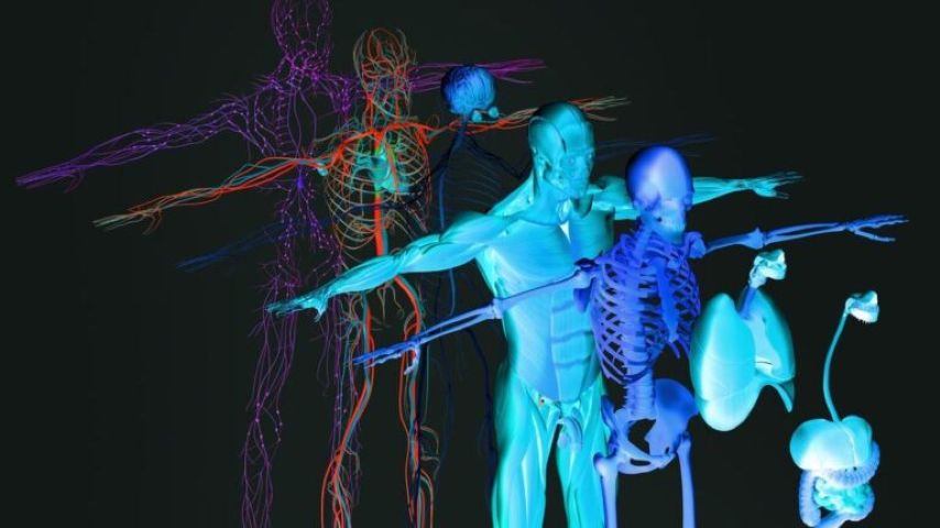
Researchers have developed a technology that combines ultrasound and photoacoustic imaging and collects images of both tissue and blood vessels at the same time.
Medical imaging is essential technology for the diagnosis and treatment of wide range of health conditions, including trauma, infection, cancer, and chronic diseases. But today’s standard imaging methods—such as magnetic resonance imaging (MRI) ultrasound, computed tomography (CT), and x-ray—have limitations, such as procedural cost, time required for each scan, how large an area can be captured, and the level of detail that can be provided.
To boost the capabilities and effectiveness of medical imaging, researchers from the Keck School of Medicine at the University of Southern California (USC) and the California Institute of Technology (Caltech) teamed up to create a non-invasive process that quickly collects 3D images of the human body. The process combines ultrasound and photoacoustic imaging (which detects sound waves generated by light) to simultaneously take images of both tissue and blood vessels.
How it works
The research effort involved combined two imaging methods, rotational ultrasound tomography (RUST) and photoacoustic tomography (PAT).
RUST directs sound waves at an area being imaged. But instead of using a single detector to create a 2D image, it uses an arc of detectors to recreate a 3D volumetric image of the body’s tissues. PAT directs a beam of laser light at the same area, which is absorbed by hemoglobin molecules in the blood. These molecules vibrate and give off ultrasonic frequencies, which are measured by the same detectors to create 3D images of blood vessels.
“We combined the power of ultrasonography for morphological assessment of soft tissue with photoacoustic tomography for visualizing blood vessels to enable three-dimensional (3D) panoramic imaging,” said Charles Liu, MD and professor of clinical neurological surgery at the Keck School of Medicine, director of the USC Neurorestoration Center, and co-leader of the research team.
You May Also Want To Read: Imaging Tool Reveals Oral Cancer Early
You May Also Want To Read: Imaging Tool Reveals Oral Cancer Early
Fast panoramic rotational RUST and PAT were integrated for hybrid rotational ultrasound and photoacoustic tomography (RUS-PAT), which obtains 3D ultrasound structural and PAT angiographic images of the human body.
“The rotational ultrasound tomography functionality is achieved using a single-element ultrasonic transducer for ultrasound transmission and rotating arc-shaped arrays for 3D panoramic detection,” said Liu. “By switching the acoustic source to a light source, the system is conveniently converted to PAT mode to acquire angiographic images in the same region.”
 The technology can capture images of both tissue structure and blood vessels across a region up to 10 centimeters wide, all in about 10 seconds. Image: Getty Images
RUS-PAT offers several potential benefits over existing medical imaging tools. For example, RUS-PAT is less expensive to build than other imaging systems, RUS-PAT avoids the radiation needed for x-ray and CT scans, and provides more sophisticated images than conventional ultrasound.
“When we think about the critical limitations of current medical imaging, including expense, field of view, spatial resolution, and time to scan, this platform addresses many of them,” Liu said.
“When we think about the critical limitations of current medical imaging, including expense, field of view, spatial resolution, and time to scan, this platform addresses many of them,” Liu said.
Impressive results
“Using RUS-PAT, we successfully imaged the human head, breast, hand, and foot with a 10-cm-diameter field of view, submillimeter isotropic resolution, and 10 s imaging time for each modality,” said co-leader Lihong Wang, professor of medical engineering and electrical engineering at Caltech. “Our research showed proof-of-concept that 3D RUS-PAT can be a powerful tool for high-speed, dual-contrast imaging of the human body with potential for rapid clinical translation.”This innovative technology has created a new way for ultrasound and photoacoustic imaging systems to work together, creating comprehensive 3-D images that allow physicians to make better-informed decisions about treatments and disease management. Another benefit is that RUS-PAT does not use ionizing radiation or strong magnets.
Ultrasound and photoacoustic imaging will likely be critical for the development of disease-specific diagnostics and therapies, especially diabetes and venous diseases. For example, RUS-PAT will provide detailed imagery to help medical teams identify at-risk limbs and inform interventions to preserve function in diabetic foot disease and other vascular conditions.
Next steps
Major research goals for the team are global implementation and affordability—rapid, low-cost imaging of the foot alone could aid millions of people living with painful diabetic foot complications.
Discover the Benefits of ASME Membership
Discover the Benefits of ASME Membership
More work is needed before RUS-PAT is ready for clinical use. One major challenge for brain application is that the human skull distorts RUS-PAT signals, making it hard to collect clear images of the brain.
The researchers are exploring novel approaches to solve this problem, including adjustments to ultrasound frequency. Further improvements are also needed to ensure consistent image quality across scans. “We are now continuing to refine the system as we move toward future clinical use,” Liu said.
Mark Crawford is a technology writer in Corrales, N.M.
The researchers are exploring novel approaches to solve this problem, including adjustments to ultrasound frequency. Further improvements are also needed to ensure consistent image quality across scans. “We are now continuing to refine the system as we move toward future clinical use,” Liu said.
Mark Crawford is a technology writer in Corrales, N.M.
Researchers have developed a technology that combines ultrasound and photoacoustic imaging and collects images of both tissue and blood vessels at the same time.

Writer and editor at MC2 Solutions
Related Content


Oct 22, 2026
Developing Talent Where Impact Happens

How the E4C Fellowship helps early-career engineers build skills in sustainable development and energy access, featuring fellows from ASU’s LEAPS lab.
Oct 21, 2026
Manufacturing Engineer Shares and Adapts

Connections are vital to create networking, training, and community engagement opportunities, along with access to critical resources.

Oct 21, 2026
ESOPs Makes Mechanical Engineers Stakeholders


Mechanical engineers are uniquely positioned to shift into fast-growing sectors.

Oct 20, 2026
The Evolution of the Plug-In Hybrid