Magnetic Beads Quickly Identify Harmful Bacteria
Magnetic Beads Quickly Identify Harmful Bacteria
Using Raman spectroscopy and immunomagnetic beads, researchers find a needle in a haystack.
When it comes to food borne illnesses, it can take days or even weeks to identify the source bacteria through laboratory testing. The food samples may only contain a few cells of bacteria, requiring scientists to grow the cells into a larger sample for detection or further examination under a microscope. With no effective way of isolating the source bacteria from the rest of the sample, scientists are searching for a needle in a haystack.
Two research labs at the Massachusetts Institute of Technology (MIT) have come together to magnetically isolate and detect bacteria in samples. Using Raman spectroscopy and immunomagnetic beads, they were able to detect the bacteria using the beads’ unique Raman optical signature. The ability to isolate and detect the bacteria means laboratory testing can be completed in hours or minutes, rather than days or weeks. When it comes to testing for multiple bacteria in a single sample, scientists can use a multiplexing method with antibody coatings attracting multiple types of bacteria or a mixture of beads targeting each type of bacteria.
The researchers have started with salmonella and E. coli samples, with the intention of detecting food borne illnesses and sepsis. Sepsis, also referred to as blood poisoning, occurs when bacteria infect the bloodstream, triggering strong immune responses and widespread inflammation. The wide-ranging symptoms and causes make sepsis hard to identify and treat. The testing process can also take days or weeks to complete, impacting the critical time for patient treatment.
Become a Member: How to Join ASME
“We envision going from the patient blood sample to being able to inform what kind of treatment is most appropriate and do so in a very quick manner” explained Rohit Karnik, Tata Professor in mechanical engineering at MIT and associate director of the Abdul Latif Jameel Water and Food Systems Lab. Karnik’s lab has been working to develop separation methods to isolate molecules from samples and improve testing methods.
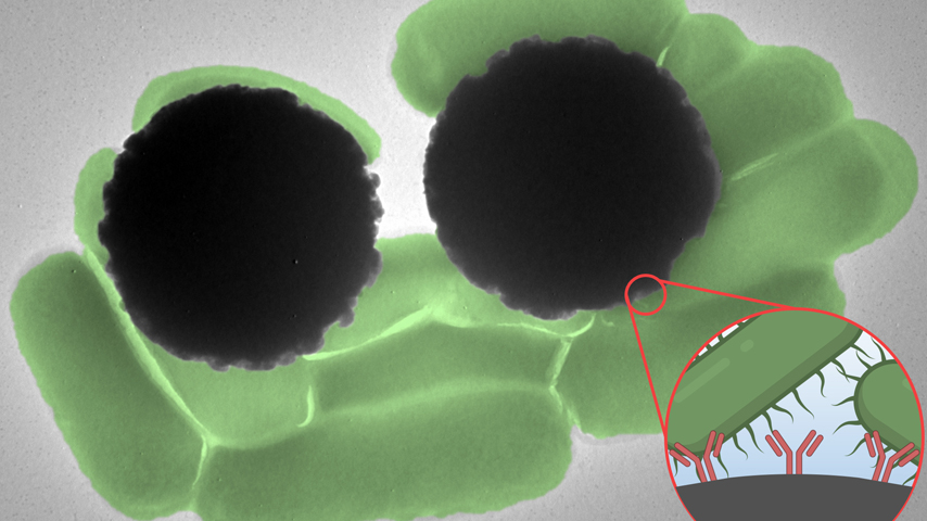
The immunomagnetic beads, ~2.5 micrometers in diameter, contain a magnetic core with a polymer shell. A salmonella antibody coating was applied to the beads before being placed in a water sample spiked with salmonella. Using the beads’ magnetic core, a magnetic field was applied to isolate the beads and allow for faster extraction of bead-bound salmonella.
Raman spectroscopy is an optical imaging technique to detect the light scattered back from a molecule or chemical compound. Using special tools, a light is shone on the molecules in the sample. Some light bounces off, while some is absorbed. The light bouncing off is called scattered light. Scientists study the unique pattern of scattered light to identify different molecules within the sample.
“The unique benefit of Raman spectroscopy is the intensity, nature, and color of light are unique to the molecular composition of the material. Similar to how a fingerprint would differentiate people around the world, this scattering serves as a fingerprint for different molecules and chemical compounds as well as bigger biological objects such as cells and in our case immunomagnetic beads” said Loza Tadesse, Brit (1961) & Alex (1949) d'Arbeloff Career Development assistant professor of mechanical engineering at MIT, and associate member of the Ragon Institute of MGH, MIT and Harvard. The Tadesse lab has been developing optical detection methods using Raman spectroscopy.
More for You: Designing a Golden Eye for the Sky
The bacteria itself often has a weak Raman signature making them hard to identify at low concentrations within large sample volumes. Contrastingly, the researchers found that the immunomagnetic beads have a strong and unique Raman signature, making them relatively easy to identify. While this does not necessarily mean they can identify the bacteria from their own Raman signature, they can isolate the beads bound to bacteria from the sample and provide a positive or negative result directly from the signature of the beads within fractions of a second.
“The immunomagnetic beads aren't enhancing the Raman spectra of the bacteria, rather we are seeing the Raman spectra of the beads themselves. They're acting as Raman reporters” said Marissa McDonald, a second-year graduate student in the Health Sciences and Technology Medical Engineering and Medical Physics program at MIT.
The speed of detection using immunomagnetic beads and Raman spectroscopy is extremely fast. When the light is applied to the sample, the Raman signature can be detected within 0.5 seconds. Additional time is needed for sample collection and preparation, but the ability to obtain results in a matter of hours or minutes after sample collection has the potential to greatly change how the medical field identifies and treats illnesses.
“We can detect the strong Raman peak signal from the immunomagnetic beads with shorter exposure time and lower laser power,” noted Jongwan Lee, a postdoctoral associate at MIT working in the Karnik lab.
The research to date was based on predefined samples with bacteria suspended in water and tested against control samples without the bacteria. This step allowed researchers to test their process in a controlled manner with a known bacteria source and no contaminants. The next step is to test their process on clinical samples for food poisoning and sepsis related applications where the bacteria is not confirmed to be present or other contaminants may exist in the sample. As they continue to test their research and push the performance limits, they can move into clinical studies and approvals to develop a portable prototype to use in medical facilities or food processing centers to identify bacteria in real time.
Nicole Imeson is an engineer and technology writer in Calgary, Alberta.
Two research labs at the Massachusetts Institute of Technology (MIT) have come together to magnetically isolate and detect bacteria in samples. Using Raman spectroscopy and immunomagnetic beads, they were able to detect the bacteria using the beads’ unique Raman optical signature. The ability to isolate and detect the bacteria means laboratory testing can be completed in hours or minutes, rather than days or weeks. When it comes to testing for multiple bacteria in a single sample, scientists can use a multiplexing method with antibody coatings attracting multiple types of bacteria or a mixture of beads targeting each type of bacteria.
The researchers have started with salmonella and E. coli samples, with the intention of detecting food borne illnesses and sepsis. Sepsis, also referred to as blood poisoning, occurs when bacteria infect the bloodstream, triggering strong immune responses and widespread inflammation. The wide-ranging symptoms and causes make sepsis hard to identify and treat. The testing process can also take days or weeks to complete, impacting the critical time for patient treatment.
Become a Member: How to Join ASME
“We envision going from the patient blood sample to being able to inform what kind of treatment is most appropriate and do so in a very quick manner” explained Rohit Karnik, Tata Professor in mechanical engineering at MIT and associate director of the Abdul Latif Jameel Water and Food Systems Lab. Karnik’s lab has been working to develop separation methods to isolate molecules from samples and improve testing methods.
The immunomagnetic beads, ~2.5 micrometers in diameter, contain a magnetic core with a polymer shell. A salmonella antibody coating was applied to the beads before being placed in a water sample spiked with salmonella. Using the beads’ magnetic core, a magnetic field was applied to isolate the beads and allow for faster extraction of bead-bound salmonella.
Raman spectroscopy is an optical imaging technique to detect the light scattered back from a molecule or chemical compound. Using special tools, a light is shone on the molecules in the sample. Some light bounces off, while some is absorbed. The light bouncing off is called scattered light. Scientists study the unique pattern of scattered light to identify different molecules within the sample.
“The unique benefit of Raman spectroscopy is the intensity, nature, and color of light are unique to the molecular composition of the material. Similar to how a fingerprint would differentiate people around the world, this scattering serves as a fingerprint for different molecules and chemical compounds as well as bigger biological objects such as cells and in our case immunomagnetic beads” said Loza Tadesse, Brit (1961) & Alex (1949) d'Arbeloff Career Development assistant professor of mechanical engineering at MIT, and associate member of the Ragon Institute of MGH, MIT and Harvard. The Tadesse lab has been developing optical detection methods using Raman spectroscopy.
More for You: Designing a Golden Eye for the Sky
The bacteria itself often has a weak Raman signature making them hard to identify at low concentrations within large sample volumes. Contrastingly, the researchers found that the immunomagnetic beads have a strong and unique Raman signature, making them relatively easy to identify. While this does not necessarily mean they can identify the bacteria from their own Raman signature, they can isolate the beads bound to bacteria from the sample and provide a positive or negative result directly from the signature of the beads within fractions of a second.
“The immunomagnetic beads aren't enhancing the Raman spectra of the bacteria, rather we are seeing the Raman spectra of the beads themselves. They're acting as Raman reporters” said Marissa McDonald, a second-year graduate student in the Health Sciences and Technology Medical Engineering and Medical Physics program at MIT.
The speed of detection using immunomagnetic beads and Raman spectroscopy is extremely fast. When the light is applied to the sample, the Raman signature can be detected within 0.5 seconds. Additional time is needed for sample collection and preparation, but the ability to obtain results in a matter of hours or minutes after sample collection has the potential to greatly change how the medical field identifies and treats illnesses.
“We can detect the strong Raman peak signal from the immunomagnetic beads with shorter exposure time and lower laser power,” noted Jongwan Lee, a postdoctoral associate at MIT working in the Karnik lab.
The research to date was based on predefined samples with bacteria suspended in water and tested against control samples without the bacteria. This step allowed researchers to test their process in a controlled manner with a known bacteria source and no contaminants. The next step is to test their process on clinical samples for food poisoning and sepsis related applications where the bacteria is not confirmed to be present or other contaminants may exist in the sample. As they continue to test their research and push the performance limits, they can move into clinical studies and approvals to develop a portable prototype to use in medical facilities or food processing centers to identify bacteria in real time.
Nicole Imeson is an engineer and technology writer in Calgary, Alberta.
Using Raman spectroscopy and immunomagnetic beads, researchers find a needle in a haystack.

Principal at Vertica Engineering and freelance writer
Related Content

Jul 24, 2026
Quiz: Extinct Classroom Engineering
Plenty of engineering innovations could be found in the classroom before computers and tablets took over. What do you remember about them?

Jul 15, 2026
Water Barrier Seeks to Protect Sailors from Jet Noise

Researchers are testing water barriers as a way to reduce the dangerous jet noise levels naval personnel experience while working on aircraft carriers.

Jul 14, 2026
A Better Step for People with Above-Knee Amputations

MIT researchers designed an affordable prosthetic foot that helps people with above-knee amputations achieve a more natural stride.

Jul 13, 2026
Exoskeleton Improves Mobility for Stroke Survivors

University of Utah researchers are piloting a 5.5-pound wearable robotic solution to help individuals with hemiparesis walk.