A Portable Alternative to Mammograms
A Portable Alternative to Mammograms
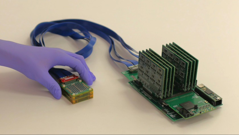
A new wearable ultrasound system generates real-time 3D breast images while reducing cost, improving comfort, and expanding access to screening.
Annual mammogram screening is considered the gold standard for detecting breast cancer. But for high-risk patients and underserved communities, mammography is far from perfect, and it presents challenges that can hinder early detection.
Even with regular screenings, cancer can be difficult to spot, as dense breast tissue and tumors are often indistinguishable in mammogram imagery.
Mammograms also rely on expensive imaging equipment and must be administered by skilled technicians in a clinic or medical facility, leaving women in underserved communities with limited access to this essential diagnostic test.
And as any woman who’s ever undergone a mammogram will tell you, it’s not the most comfortable experience, as each breast gets pressed between two plates to spread out tissues for imaging.
A research team from the Massachusetts Institute of Technology (MIT) has miniaturized a portable, low-cost ultrasound device they innovated in 2023 to address each of those challenges, improving visualization of breast abnormalities while potentially reducing the need for some in-clinic imaging appointments.
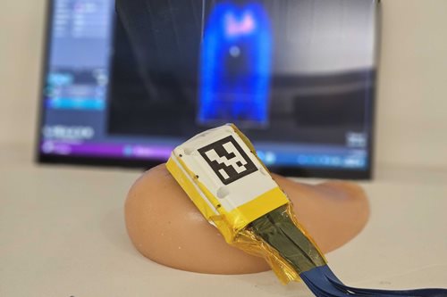 A 3D ultrasound probe is tested on a breast tissue phantom containing an embedded tumor model. Image: MIT
Previously, the team developed a honeycomb-shaped device consisting of an array of ultrasound transducers that were incorporated into a flexible patch that could attach to a bra. The design enabled the wearer to move the tracker along the patch to capture images of the breast from different angles.
A 3D ultrasound probe is tested on a breast tissue phantom containing an embedded tumor model. Image: MIT
Previously, the team developed a honeycomb-shaped device consisting of an array of ultrasound transducers that were incorporated into a flexible patch that could attach to a bra. The design enabled the wearer to move the tracker along the patch to capture images of the breast from different angles.
But because the device was limited to two-dimensional imagery, gaps in coverage were possible. The 2D images could be combined to generate a 3D representation of the breast tissue, but small abnormalities could still be missed. And to view the images, the array needed to be connected to a traditional, costly machine about the size of a refrigerator.
With their latest research, the team modified their original design to produce a more compact, fully portable ultrasound array capable of generating a 3D image of the entire breast from just two or three locations.
Roughly the size of a smartphone, the new device consists of an ultrasound probe attached to a motherboard that processes the data. The system can connect to a laptop to display wide-angle, real-time 3D images.
“Instead of massive machines that use radiation, our device uses miniaturized ultrasound sensors housed in a wearable patch,” said Canan Dagdeviren, the study’s senior author and an associate professor of media arts and sciences at MIT. “It connects to a small, portable data system, making it easy to deploy anywhere.”
This work was recently published as “Real-Time 3D Ultrasound Imaging with an Ultra-Sparse, Low Power Architecture” in the journal Advanced Healthcare Materials.
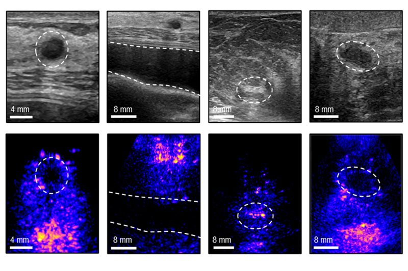 Four in vivo ultrasound images show, from left to right, a cyst, breast implant, dense fibrous tissue, and solid mass. Conventional 2D ultrasound images appear in the top row, while the corresponding 3D images are shown below. Image: MIT
Because mammograms capture two-dimensional X-ray images, dense breast tissue and tumors often show up in imaging as the exact same white color. Traditional ultrasounds can solve this problem, but they require a highly trained specialist to manually scan the breast for 20 to 30 minutes. “Needing a specialist drives up the cost significantly and gates accessibility for many patients,” Dagdeviren explained.
Four in vivo ultrasound images show, from left to right, a cyst, breast implant, dense fibrous tissue, and solid mass. Conventional 2D ultrasound images appear in the top row, while the corresponding 3D images are shown below. Image: MIT
Because mammograms capture two-dimensional X-ray images, dense breast tissue and tumors often show up in imaging as the exact same white color. Traditional ultrasounds can solve this problem, but they require a highly trained specialist to manually scan the breast for 20 to 30 minutes. “Needing a specialist drives up the cost significantly and gates accessibility for many patients,” Dagdeviren explained.
By capturing a wide-angle 3D image of the breast, the device eliminates the need for a specialist to perform the scan.
“Our device takes the deep tissue visibility of an ultrasound and automates it,” Dagdeviren said. “Patient comfort is crucial for screening compliance. Our technology is designed as a soft, conformable patch that rests naturally on the body.”
Making the ultrasound system portable presented a primary challenge for researchers, as 3D imaging usually requires massive electronic machinery. Thorough imaging also requires sufficient acoustic power to see deep enough into the human body. “We overcame this by completely redesigning how the ultrasound signals are sent and processed. This drastically lowered the power and space needed while maintaining crystal clear 3D images,” Dagdeviren said.
 A close-up view shows the chip's wire-bonded sensor array architecture. Image: MIT
To engineer a miniaturized ultrasound system, the research team relied on advanced engineering concepts to dramatically reduce the number of sensors the device needed.
A close-up view shows the chip's wire-bonded sensor array architecture. Image: MIT
To engineer a miniaturized ultrasound system, the research team relied on advanced engineering concepts to dramatically reduce the number of sensors the device needed.
One approach they used was sparse array imaging, an ultrasound technique used to generate high-resolution images with a reduced number of components, enabling a faster process and less expensive equipment.
The team also incorporated compressed sensing, a technique for acquiring and reconstructing images from a limited number of samples. By rethinking those limits, researchers were able to capture a full 3D image with a fraction of the usual hardware. “This allowed us to shrink the physical patch on the patient’s body to a remarkably small footprint,” Dagdeviren said.
Researchers tested the device on a 71-year-old woman with a history of breast cysts. Capable of imaging up to 15 centimeters deep, the device successfully produced accurate images of the cysts and generated a 3D view of the surrounding breast tissue.
“We demonstrated that our portable infrastructure could clearly detect and differentiate both solid tumors and fluid-filled cysts in human breast tissue. We were also able to identify microcalcifications, matching the capabilities of large hospital machines,” Dagdeviren said.
Researchers are actively spinning the technology out of MIT into a medical device startup to commercialize the innovation, working through the National Science Foundation’s Innovation Corps (I-Corps) program to pursue pathways to U.S. Food and Drug Administration approval.
“In the near term, we want to equip mobile clinics globally with this device so no patient is turned away because they have dense breasts,” Dagdeviren said.
More For You: Fueling Immunity from Within
Although mammography would be a difficult space to enter or replace in the United States because of well-established practices and infrastructure, Dagdeviren said the portable ultrasound device could serve as a powerful complementary tool to fill in the gaps for dense breast screening. And in medically underserved regions globally, the device could even be the primary screening tool for breast cancer. “A large part of the world has absolutely no access to mammogram machines,” Dagdeviren said.
By solving for the core problem of making a 3D ultrasound wearable and low-power, Dagdeviren said the technology could be easily adapted to monitor cardiovascular health, organ function, and even fetal movement during pregnancy without the need for a clinic visit.
Within the decade, the research team envisions widespread use of the device for mass screenings in developing countries and in the homes of high-risk women. “This will enable safe, automated, and frequent breast cancer monitoring right from their living rooms,” Dagdeviren said.
Autumn Giusti is an independent writer in New Orleans.
Even with regular screenings, cancer can be difficult to spot, as dense breast tissue and tumors are often indistinguishable in mammogram imagery.
Mammograms also rely on expensive imaging equipment and must be administered by skilled technicians in a clinic or medical facility, leaving women in underserved communities with limited access to this essential diagnostic test.
And as any woman who’s ever undergone a mammogram will tell you, it’s not the most comfortable experience, as each breast gets pressed between two plates to spread out tissues for imaging.
A research team from the Massachusetts Institute of Technology (MIT) has miniaturized a portable, low-cost ultrasound device they innovated in 2023 to address each of those challenges, improving visualization of breast abnormalities while potentially reducing the need for some in-clinic imaging appointments.
A 3D ultrasound probe is tested on a breast tissue phantom containing an embedded tumor model. Image: MIT
But because the device was limited to two-dimensional imagery, gaps in coverage were possible. The 2D images could be combined to generate a 3D representation of the breast tissue, but small abnormalities could still be missed. And to view the images, the array needed to be connected to a traditional, costly machine about the size of a refrigerator.
With their latest research, the team modified their original design to produce a more compact, fully portable ultrasound array capable of generating a 3D image of the entire breast from just two or three locations.
Roughly the size of a smartphone, the new device consists of an ultrasound probe attached to a motherboard that processes the data. The system can connect to a laptop to display wide-angle, real-time 3D images.
“Instead of massive machines that use radiation, our device uses miniaturized ultrasound sensors housed in a wearable patch,” said Canan Dagdeviren, the study’s senior author and an associate professor of media arts and sciences at MIT. “It connects to a small, portable data system, making it easy to deploy anywhere.”
This work was recently published as “Real-Time 3D Ultrasound Imaging with an Ultra-Sparse, Low Power Architecture” in the journal Advanced Healthcare Materials.
Delivering on a promise
The development of the device has been more than a decade in the making for Dagdeviren, whose aunt was diagnosed with late-stage breast cancer in 2015. Despite regular mammograms, Dagdeviren’s aunt’s tumors had been masked by dense breast tissue. Sitting at her aunt’s bedside, Dagdeviren sketched the initial concept for a wearable ultrasound that would specifically address the challenge of breast tissue density. “I promised her I would make it a reality,” she said.
Four in vivo ultrasound images show, from left to right, a cyst, breast implant, dense fibrous tissue, and solid mass. Conventional 2D ultrasound images appear in the top row, while the corresponding 3D images are shown below. Image: MIT
By capturing a wide-angle 3D image of the breast, the device eliminates the need for a specialist to perform the scan.
“Our device takes the deep tissue visibility of an ultrasound and automates it,” Dagdeviren said. “Patient comfort is crucial for screening compliance. Our technology is designed as a soft, conformable patch that rests naturally on the body.”
Shrinking by rethinking
Making the ultrasound system portable presented a primary challenge for researchers, as 3D imaging usually requires massive electronic machinery. Thorough imaging also requires sufficient acoustic power to see deep enough into the human body. “We overcame this by completely redesigning how the ultrasound signals are sent and processed. This drastically lowered the power and space needed while maintaining crystal clear 3D images,” Dagdeviren said.
A close-up view shows the chip's wire-bonded sensor array architecture. Image: MIT
One approach they used was sparse array imaging, an ultrasound technique used to generate high-resolution images with a reduced number of components, enabling a faster process and less expensive equipment.
The team also incorporated compressed sensing, a technique for acquiring and reconstructing images from a limited number of samples. By rethinking those limits, researchers were able to capture a full 3D image with a fraction of the usual hardware. “This allowed us to shrink the physical patch on the patient’s body to a remarkably small footprint,” Dagdeviren said.
Researchers tested the device on a 71-year-old woman with a history of breast cysts. Capable of imaging up to 15 centimeters deep, the device successfully produced accurate images of the cysts and generated a 3D view of the surrounding breast tissue.
“We demonstrated that our portable infrastructure could clearly detect and differentiate both solid tumors and fluid-filled cysts in human breast tissue. We were also able to identify microcalcifications, matching the capabilities of large hospital machines,” Dagdeviren said.
Filling in the gaps
Researchers are actively spinning the technology out of MIT into a medical device startup to commercialize the innovation, working through the National Science Foundation’s Innovation Corps (I-Corps) program to pursue pathways to U.S. Food and Drug Administration approval. “In the near term, we want to equip mobile clinics globally with this device so no patient is turned away because they have dense breasts,” Dagdeviren said.
More For You: Fueling Immunity from Within
Although mammography would be a difficult space to enter or replace in the United States because of well-established practices and infrastructure, Dagdeviren said the portable ultrasound device could serve as a powerful complementary tool to fill in the gaps for dense breast screening. And in medically underserved regions globally, the device could even be the primary screening tool for breast cancer. “A large part of the world has absolutely no access to mammogram machines,” Dagdeviren said.
By solving for the core problem of making a 3D ultrasound wearable and low-power, Dagdeviren said the technology could be easily adapted to monitor cardiovascular health, organ function, and even fetal movement during pregnancy without the need for a clinic visit.
Within the decade, the research team envisions widespread use of the device for mass screenings in developing countries and in the homes of high-risk women. “This will enable safe, automated, and frequent breast cancer monitoring right from their living rooms,” Dagdeviren said.
Autumn Giusti is an independent writer in New Orleans.
A new wearable ultrasound system generates real-time 3D breast images while reducing cost, improving comfort, and expanding access to screening.

Independent writer, editor, and content specialist.
Related Content

Aug 20, 2026
Artificial Muscles That Move to Sound

Microbubbles of varying sizes embedded in polymers have different acoustic responses, which can help move artificial muscles made from this material.


Jul 24, 2026
Quiz: Extinct Classroom Engineering

Plenty of engineering innovations could be found in the classroom before computers and tablets took over. What do you remember about them?
Jul 14, 2026
A Better Step for People with Above-Knee Amputations

MIT researchers designed an affordable prosthetic foot that helps people with above-knee amputations achieve a more natural stride.
Jul 13, 2026
Exoskeleton Improves Mobility for Stroke Survivors

University of Utah researchers are piloting a 5.5-pound wearable robotic solution to help individuals with hemiparesis walk.