A Road Map for the Spinal Cord
A Road Map for the Spinal Cord
A multi-institutional research effort seeks to improve a flexible probe system that will uncover how signals travel the spinal cord, holding promise for improved therapies.
Researchers at Rice University in Houston are leading a multi-institutional research project that aims to optimize nanoelectronic threads (NETs) for use in the spinal cord.
Nearly a decade ago, scientists developed NETs to record electronic signals in the brain. The process begins by strategically placing NETs in a test subject’s brain and connecting them to an external device, such as a computer. Small electronic signals can then be measured and recorded as the subject completes specific movements, allowing researchers to better understand how data transfers within the brain to other parts of the body. Through various studies and tests, the research team built a roadmap of the brain’s electronic signals and correlated actions.
Now, a 4-year, $6.25-million grant from the National Institute of Health will help the research team to do the same for the spinal cord.
Spinal cord construction
In the outer layer of the brain, neurons, or white matter, transfer electronic signals. Located in the brain’s internal layers are nerve fibers, or grey matter. The spinal cord is the opposite of the brain: Nerve fibers are on the outside, protecting neurons on the inside. But current spinal cord testing methods measure electronic signals on the surface of the cord, which does not adequately capture the electronic signals of the neurons inside.
 Principal investigator Chong Xie (left) is an associate professor of electrical and computer engineering and neuroengineering at Rice University. Lan Luan is co-investigator and an assistant professor of electrical and computer engineering. Photo: Gustavo Raskosky/Rice University
Become a Member: How to Join ASME
Rice is one of four major institutions involved in the grant project hoping to answer several of these physiological questions. The others are the Salk Institute for Biological Studies in southern California, Carnegie Mellon University in Pittsburgh, and the University of California, San Francisco.
In addition to understanding how electric signals generate motion patterns and how the spinal cord functions, scientists are hoping to understand pain circuits. It is believed pain signals from various parts of the body pass through the spinal cord on their way to the brain to be registered. Intercepting those signals at the spinal cord as an alternative pain management technique could offer relief to patients living with chronic pain.
Nanoelectronic threads
The spinal cord is a more mobile organ than the brain, requiring more flexibility and range of motion with the test apparatus. “No one has recorded individual neurons when the subject's moving. If you want to follow something moving, the device itself must be moving in a flexible way as well,” Xie said.
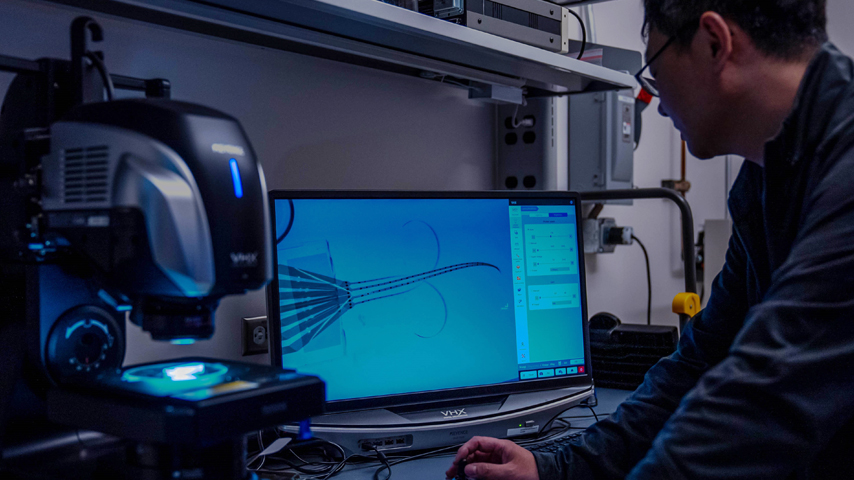
This presented a challenge, as current test apparatuses are rigid and cumbersome. NETs are thin, flexible polymers with a low bending stiffness making them a good candidate to place within the mobile and flexible spinal cord. Embedding NETs in the spinal cord, researchers can record individual neuron activity, without affecting the surrounding tissue and with no scar tissue or neuronal degeneration after implantation.
But implanting NETs can also be challenging due to their flexibility, much like pushing a rope. When testing the brain, a test subject undergoes a craniotomy, temporarily removing a segment of the skull, to expose the brain where the NETs will be attached. Scientists use a rigid microwire, referred to as an insertion shuttle, and bio-dissolvable adhesive to secure NETs during insertion. However, there is a small window during which the NETs can be placed, before cerebrospinal fluid dissolves the adhesive and the shuttle is retracted. A similar procedure is used to implant NETs in the spinal cord.
You Might Also Enjoy: Electronic Skin Sends Sensations to the Brain
After the subject recovers from surgery, researchers can monitor the subject’s exact motion and correlate the neural activity that the NETs register to build a road map of electronic signals through the spinal cord. Rice researchers are currently testing NETs in the spinal cords of mice using a gate and treadmill set up.
One limitation with current NETs is they can only carry 32 electrode channels, which is not enough to record adequate spinal cord data.
“This is an engineering task to scale things up in size so that we can work with larger spinal cords. Currently, we only work with rodents or mice,” Xie explained. “We are hoping to eventually work with rats, non-human primates, and eventually with humans.”
One of the grant’s major goals is to scale NETs to allow upward of 512 electrode channels to record even more electronic signals, which will help researchers gain a far better understanding of spinal cord functions.
Nicole Imeson is an engineer and technology writer in Calgary, Alberta.
A multi-institutional research effort seeks to improve a flexible probe system that will uncover how signals travel the spinal cord, holding promise for improved therapies.

Principal at Vertica Engineering and freelance writer
Related Content


