3D Modeling from the Womb
3D Modeling from the Womb
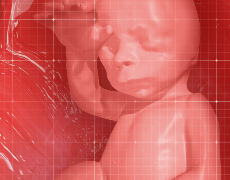
New technology transforms MRI and ultrasound data into a 3D virtual reality model of a fetus. Fetus image: RSNA
These days parents can get to know their babies pretty early on thanks to ultrasounds that let them have a peek at the fetus long before birth. But the view they offer is always from the outside looking in. Now researchers at the Federal University of Rio de Janeiro are giving parents and doctors a view from within the womb using virtual reality.
Dr. Heron Werner, an OBGYN and professor of biotechnology at the university, has long worked to bring 3D representations of what was going on in utero to the outside. In 2008 he started working on creating better 3D images of fetuses so they could be printed out and handed to doctors. “The main objective in the beginning was to prepare difficult pathologies and print them for discussion,” he says.
Now Werner and his colleagues are combining their imaging techniques with Oculus Rift goggles to allow parents to climb right into the uterus and get acquainted with their offspring.
The trick is the combination of ultrasound and MRI. Though ultrasound imaging has gotten better and better over the years, the field of view it offers is fairly narrow. At any one moment it can show a hand, or a foot, or an ear, but not the whole body. With the MRI, you get the whole picture. But, thanks to long the duration of the scan, there are often motion artifacts.
To minimize fetal and maternal activity, Werner asks mothers to hold their breath for twenty seconds. “Sometimes you have to try again and again and again.” Occasionally, when breath holding is not quite sufficient to calm all fidgeting on the part of the baby or the mother, Werner will offer a little light sedation.
Combining the MRI with the ultrasound gives a very detailed composite image. “If I didn’t get the right ear with the MRI, I can get it from the ultrasound and put it together,” says Werner.
The putting together is chief challenge of the project. “The most difficult thing was to find the best software to combine them,” says Jorge Lopes, an industrial designer who worked on the programming side of the project, “and working with the outputs for different 3D printing machines, and the virtual reality—they all work with different outputs.”
With those problems largely solved, the current obstacle to widespread use of the technology is the time it takes. Not only do the scans take from twenty minutes to five hours, but the images must be manually assembled and cleaned. “There is no retouching as in Photoshop,” says Lopes. “We just have to find the biomechanical points.”
Though parents-to-be may ooh and ahh over the close encounter with their little one, the virtual reality goggles offer more than a preview of cuteness. MRI and ultrasound capture more than the surface of things, after all. Doctors and parents can now explore the baby in a way that would be impossible after birth. They can zoom through the entire anatomy, travel along arteries, inspect the heart, wander around the brain; an incredible advantage should there be any pathologies. If there are tumors in an airway, for instance, a surgeon can find out exactly where they sit, and even practice a surgery before birth. Alternatively, the passageway could be printed out and the surgeon could plan his best moves on a model.
A 3D printed fetus would also allow blind parents to “touch the image,” as Werner puts it, giving them, for the first time in history, a tactile view of a baby inside the womb.
Werner’s imaging process needn’t be limited to sneak peeks of babies. “The same process can be used to navigate inside the uterus and to study the fallopian tubes,” says Werner. “The aim here is to see fibromas, polyps, et cetera.”
Unless scanning technology changes wildly we’re not likely to view babies in motion before they are born anytime soon. And until we find a way to get a pair of VR goggles to a baby in the womb, the interaction will remain one-way.
Michael Abrams is an independent writer.
The main objective in the beginning was to prepare difficult pathologies and print them for discussion.Dr. Heron Werner, Federal University of Rio de Janeiro
New technology transforms MRI and ultrasound data into a 3D virtual reality model of a fetus. Fetus image: RSNA

Freelance writer based in New Jersey
Related Content


