Tiny, Dissolvable Pacemaker May Be Lifesaving for Babies
Tiny, Dissolvable Pacemaker May Be Lifesaving for Babies
Researchers have developed a pacemaker small enough to fit inside the tip of a syringe and be non-invasively injected into the body. When it is no longer needed, it simply dissolves.
About 1 percent of all babies worldwide are born with a congenital heart defect, and about 25 percent of those babies need open-heart surgery during their first year of life. These tiny patients were the primary motivation for Northwestern University engineers to create a miniature pacemaker.
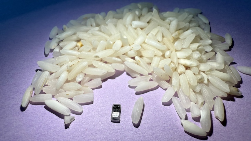
This new pacemaker — which is smaller than a grain of rice — can be fitted inside the tip of a syringe and non-invasively injected into the body. It can work for all patients, but the pacemaker was specifically designed for the tiny, fragile hearts of newborn babies with congenital heart defects.
 New tiny pacemaker (far right) next to a traditional pacemaker (left) and leadless pacemaker (center). Image: Northwestern University
“After a baby has surgery to correct the anatomical heart defect, there is a high chance of developing an electrical abnormality,” said Igor Efimov, a Northwestern experimental cardiologist who co-led the study, recently published in the journal Nature. “Fortunately, this electric dysfunction is often temporary, but for about a week, pediatric patients need a temporary pacemaker.”
New tiny pacemaker (far right) next to a traditional pacemaker (left) and leadless pacemaker (center). Image: Northwestern University
“After a baby has surgery to correct the anatomical heart defect, there is a high chance of developing an electrical abnormality,” said Igor Efimov, a Northwestern experimental cardiologist who co-led the study, recently published in the journal Nature. “Fortunately, this electric dysfunction is often temporary, but for about a week, pediatric patients need a temporary pacemaker.”
With current temporary pacemakers, a wire connects the heart to an external pulse generator through an incision in the skin. Following the recovery of normal electrical excitation, the wire is removed by pulling it out of the tissue.
“This procedure is risky,” sometimes even resulting in death, Efimov said. “Potential complications include infection, dislodgement, torn or damaged tissues, bleeding, and blood clots.”
Relevant Reads: Stamp-Like Device Treats Heart Disease, Then Dissolves
The new pacemaker addresses this issue with its biocompatible design. When it is no longer needed, it simply dissolves, eliminating the need for surgical extraction and making it much safer for the patient.
The tiny pacemaker is paired with a small wireless wearable device that mounts onto the patient’s chest to control pacing. When the device detects an irregular heartbeat, it automatically shines a light pulse to activate the pacemaker. These short pulses, which penetrate the patient’s skin, breastbone, and muscles, control the pacing.
Efimov and Northwestern bioelectronics pioneer John A. Rogers, who co-led the study, were known for their work developing the first dissolvable pacemaker for patients who need temporary pacing.
“We were approached by cardiac surgeons,” Rogers said. “They asked whether we could develop a fully wireless implantable device as a temporary pacemaker. Their motivation was for a device that could eliminate the [wires] and the need for the surgical removal—both of which expose the patients to risk.”
“The result is this tiny pacemaker, referred to as the milli-pacemaker because it’s a millimeter scale device," Efimov said.
“We have developed what is, to our knowledge, the world’s smallest pacemaker,” Rogers added. “We designed the device [with] all of the specific clinical requirements for pacing and operational timeframe, but in a fully implantable, wireless and bioresorbable format. We drew upon our previous experience in materials and microfabrication techniques for the circuits, and we introduced a new component—a light-controlled switching element—to allow for an optical scheme for controlling the operation of the device.”
In addition to making it wireless and dissolvable, cardiac surgeons requested that the researchers make it smaller, so it could be used in the smallest patients, which was challenging because their original pacemaker delivered stimulation using radio frequency. Overall, the size of the receiver antenna limited the ability to miniaturize the device.
“Instead of using the radio frequency scheme for wireless control, we developed a light-based scheme for turning the pacemaker on and delivering stimulation pulses to the surface of the heart. We used a phototransistor operating at near infrared wavelengths for good penetration through the tissue from a skin-mounted light source,” Rogers said. “This is one feature that allowed us to dramatically reduce the size.”
Discover the Benefits of ASME Membership
The power supply was another obstacle. The original device used near-field communication, requiring a built-in antenna. For the milli-pacemaker, a pair of metal electrodes was used to form a galvanic cell—a simple battery that converts chemical energy into electrical energy. When in contact with surrounding biofluids, the electrodes form a battery. The resulting chemical reactions cause the electrical current to flow, stimulating the heart.
“Testing of the milli-pacemaker has gone well so far,” said Efimov said. “We have demonstrated the function of our pacemaker and wearable device, which controls it in several models: perfused human and pig heart, in vivo in rat, and in vivo in dog. Now the goal is to show its safety and function in patients, adult and pediatric. This is challenging and will take several years.”
In addition to eventually using the milli-pacemaker in heart patients, researchers see other potential applications.
“This approach will be replicated in many other settings requiring electrical stimulation, including brain, spinal cord, autonomic nerves, gut, muscle, and many other mechanically and electrically active tissues,” Efimov said.
Rogers agreed: “These tiny pacemakers can be used more generally as light-controlled stimulators—suitable also for use with the peripheral nerves, the spinal cord, and the brain. We are looking specifically at opportunities in temporary pacing of the vagus nerve and in electrical blocking of pain signals in the peripheral nerves.”
Claudia Hoffacker is an independent writer in Minneapolis.
This new pacemaker — which is smaller than a grain of rice — can be fitted inside the tip of a syringe and non-invasively injected into the body. It can work for all patients, but the pacemaker was specifically designed for the tiny, fragile hearts of newborn babies with congenital heart defects.
New tiny pacemaker (far right) next to a traditional pacemaker (left) and leadless pacemaker (center). Image: Northwestern University
With current temporary pacemakers, a wire connects the heart to an external pulse generator through an incision in the skin. Following the recovery of normal electrical excitation, the wire is removed by pulling it out of the tissue.
“This procedure is risky,” sometimes even resulting in death, Efimov said. “Potential complications include infection, dislodgement, torn or damaged tissues, bleeding, and blood clots.”
Relevant Reads: Stamp-Like Device Treats Heart Disease, Then Dissolves
The new pacemaker addresses this issue with its biocompatible design. When it is no longer needed, it simply dissolves, eliminating the need for surgical extraction and making it much safer for the patient.
The tiny pacemaker is paired with a small wireless wearable device that mounts onto the patient’s chest to control pacing. When the device detects an irregular heartbeat, it automatically shines a light pulse to activate the pacemaker. These short pulses, which penetrate the patient’s skin, breastbone, and muscles, control the pacing.
Efimov and Northwestern bioelectronics pioneer John A. Rogers, who co-led the study, were known for their work developing the first dissolvable pacemaker for patients who need temporary pacing.
“We were approached by cardiac surgeons,” Rogers said. “They asked whether we could develop a fully wireless implantable device as a temporary pacemaker. Their motivation was for a device that could eliminate the [wires] and the need for the surgical removal—both of which expose the patients to risk.”
“The result is this tiny pacemaker, referred to as the milli-pacemaker because it’s a millimeter scale device," Efimov said.
“We have developed what is, to our knowledge, the world’s smallest pacemaker,” Rogers added. “We designed the device [with] all of the specific clinical requirements for pacing and operational timeframe, but in a fully implantable, wireless and bioresorbable format. We drew upon our previous experience in materials and microfabrication techniques for the circuits, and we introduced a new component—a light-controlled switching element—to allow for an optical scheme for controlling the operation of the device.”
In addition to making it wireless and dissolvable, cardiac surgeons requested that the researchers make it smaller, so it could be used in the smallest patients, which was challenging because their original pacemaker delivered stimulation using radio frequency. Overall, the size of the receiver antenna limited the ability to miniaturize the device.
“Instead of using the radio frequency scheme for wireless control, we developed a light-based scheme for turning the pacemaker on and delivering stimulation pulses to the surface of the heart. We used a phototransistor operating at near infrared wavelengths for good penetration through the tissue from a skin-mounted light source,” Rogers said. “This is one feature that allowed us to dramatically reduce the size.”
Discover the Benefits of ASME Membership
The power supply was another obstacle. The original device used near-field communication, requiring a built-in antenna. For the milli-pacemaker, a pair of metal electrodes was used to form a galvanic cell—a simple battery that converts chemical energy into electrical energy. When in contact with surrounding biofluids, the electrodes form a battery. The resulting chemical reactions cause the electrical current to flow, stimulating the heart.
“Testing of the milli-pacemaker has gone well so far,” said Efimov said. “We have demonstrated the function of our pacemaker and wearable device, which controls it in several models: perfused human and pig heart, in vivo in rat, and in vivo in dog. Now the goal is to show its safety and function in patients, adult and pediatric. This is challenging and will take several years.”
In addition to eventually using the milli-pacemaker in heart patients, researchers see other potential applications.
“This approach will be replicated in many other settings requiring electrical stimulation, including brain, spinal cord, autonomic nerves, gut, muscle, and many other mechanically and electrically active tissues,” Efimov said.
Rogers agreed: “These tiny pacemakers can be used more generally as light-controlled stimulators—suitable also for use with the peripheral nerves, the spinal cord, and the brain. We are looking specifically at opportunities in temporary pacing of the vagus nerve and in electrical blocking of pain signals in the peripheral nerves.”
Claudia Hoffacker is an independent writer in Minneapolis.
Researchers have developed a pacemaker small enough to fit inside the tip of a syringe and be non-invasively injected into the body. When it is no longer needed, it simply dissolves.

Independent writer and editor
Related Content

Jul 14, 2026
A Better Step for People with Above-Knee Amputations

MIT researchers designed an affordable prosthetic foot that helps people with above-knee amputations achieve a more natural stride.

Jul 13, 2026
Exoskeleton Improves Mobility for Stroke Survivors

University of Utah researchers are piloting a 5.5-pound wearable robotic solution to help individuals with hemiparesis walk.

Jul 9, 2026
A Portable Alternative to Mammograms

A new wearable ultrasound system generates real-time 3D breast images while reducing cost, improving comfort, and expanding access to screening.

Jun 25, 2026
A Mighty Wind Project
You probably don’t know how much you fart. And turns out, nobody else does either. Researchers at the University of Maryland have created a tool to finally answer this vital question.