A Tiny Top for Tiny Brains
A Tiny Top for Tiny Brains
Researchers have essentially created the world’s tiniest EEG cap for the world’s tiniest brains to better test how cells do their thing in an organ.
A good chunk of our biological understanding comes from looking at cell cultures under a microscope. Those cultures have always been essentially two dimensional, usually at the bottom of a Petri dish. For decades that’s how we’ve grown them, and that’s how we’ve looked at them.
Human bodies, though, aren’t usually flat. Nor are the organs found within in them. To better test how cells do their thing in an organ in all three dimensions, cell biologists have, as of recent, been creating little clumps of cells they call organoids. These minuscule bits of hearts, brains, livers, lungs, etc., allow researchers to probe their workings without procuring full-sized organs the old-fashioned way—in animal or human.
But our ability to grow tiny organs has far outpaced our ability to monitor what they’re doing. We just don’t have the tools.
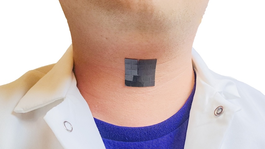
 Organoids are sensitive to pressure, so Gracias created a snuggly fitting cap made of polymer that bends when it comes in contact with water.
Those sensors work well for a culture in two dimensions, but are less than ideal for three. “If you take a flat plate and put it under a sphere, it only touches—and records activity from—a fraction of the area of the organoid,” said Gracias. To get a picture of what’s going on inside a tiny brain, you need to look at it from all sides.
Now Gracias and his colleagues have created what is essentially the world’s tiniest EEG cap for the world’s tiniest brains. In a paper he coauthored, “Shell Microelectrode Arrays (MEAs) For Brain Organoids,” which appeared in Science Advances this August, he lays out how his self-folding sensor works to cradle an organoid brain on three sides.
More Like This: Mini Kidney on a 3D-printed Chip
The size of organoid brains is only one of the difficulties that face anyone trying to read what might be going on inside. The tiny brains are as sensitive to pressure as the macro-sized version, so the cap Gracias set out to create had to fit snuggly but without crushing the brain cells. It also had to sport enough electrodes to make it useful, all within a few hundred microns.
“Brain organoids are living things—they’re fragile, you can’t use rigid materials, you can’t heat them, and they’re sensitive to toxic chemicals,” said Gracias, who had to find a material that was biocompatible and wasn’t too stiff, and use a low temperature process for fabrication and operation.
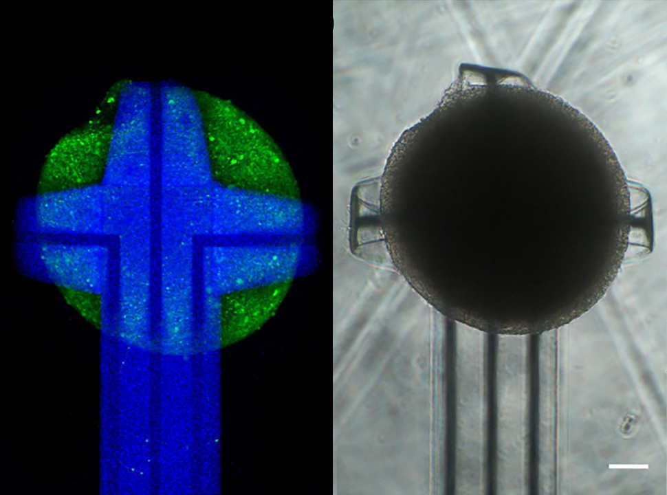
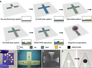
His solution was to turn to the world of self-folding origami. He used a polymer that bends when it comes in contact water, cross-linked with a conductive polymer, and cut them into the shape of a cross. When they come in contact with water, the two polymers swell differently.
Related Reading: A Pressures Sensor That’s Sensitive Under Pressure
The result is that the cross curls up on three sides, wrapping itself around whatever object is sitting at its center and cradling it with just the right amount of pressure. Mounted with three to nine electrodes, the sensor cap makes contact with the brain it holds in three dimensions.
Gracias showed not just that such an idea was feasible, but that it could easily be mass produced and in different sizes. “Just like cap sizes: small, medium large,” he said.
The potential applications for a brain organoid matched with the right instrumentation are many. For one thing, researchers could measure the effects of potentially neurotoxic chemicals on the brain, providing an alternative to animal testing. Potential drugs could also be screened without testing them on real people. And, with Gracias’ method, brain organoids could be a part of tomorrow's super computers.
You Might Also Like: Superalloy Rescues MEMS Sensors
“It’s well known that the human brain is very energy efficient and can solve problems conventional computers struggle with—face recognition and image processing,” said Gracias. “Maybe we can connect brain organoids and use them as computational elements on a chip.”
But there’s plenty of work to be done before that happens, primarily in increasing the number of cells that the little cap can measure. “The holy grail, for the whole community, is to record from every cell in the organoid,” said Gracias. “Then you get maximal information.”
Michael Abrams is a science and technology writer in Westfield, N.J.
Related Content