Biotechnology Anticipates 4D Printing
Biotechnology Anticipates 4D Printing

4D printing solutions are just beginning their evolution.
Rapid advancements in 3D printing that have fueled the development of advanced manufacturing applications are well-known. New printing techniques and their ability to print objects from a growing variety of materials such as plastics, metals, ceramics, and more allow developers and manufacturers to speed prototyping, streamline supply chains, and produce complex designs not previously possible. Even so, there are limits to what can be done because the materials are rigid.
What is the next step? It's 4D printing, a term that is a bit of a misnomer because it still relies on 3D printers. Time is the element that pushes 3D to 4D, creating printed materials that change their shape over time. “You first have to have 3D manufacturing capability and then you add smart materials,” said Howon Lee, an associate professor of mechanical and space engineering at Rutgers University.
Smart materials have the ability to change shape over time, creating a wide-ranging universe of potential new products. NASA has already produced woven metal fabrics that change shape and are foldable. Their potential use ranges from shielding a spacecraft from extreme temperatures to erecting an antenna in space. Airbus is investigating 4D-printed components that could lighten the weight and improve the performance of aircraft.
4D-printed healthcare applications may be the closest to commercial applications. Shape-shifting materials could be used for small, implantable medical devices, said Lee. Tiny, soft devices could be inserted or implanted in people, and harden when they reach the affected area. But with a few possible exceptions, wide-scale application remains years away. Most activity now is still in research and development. The market is in its infancy.
Latest in News: How Coronavirus is Reshaping the Engineering World
One survey by ResearchandMarkets.com puts the overall market for 4D printing in healthcare at a meager $9 million in 2021, rising to $32 million by 2026. In 2021, the medical and research models segment is expected to account for the largest share of the overall healthcare market. “The ability of the 4D printing technology in manufacturing smart medical models will bring significant transformation in the medical field and will support the growth of this segment in the forecast period,” according to the report. Yet, despite its small market size, emerging research shows that 4D printing could provide physicians and surgeons with new tools for everything from generating replacement skin to creating objects that respond to changes in their environment.
One example is work taking place at the Self-Assembly Lab at the Massachusetts Institute of Technology. Using Autodesk’s Cyborg software and Stratasys 3D printers, it has been developing materials for more than five years that change shape when triggered by water.
 MIT's Self-Assembly Lab employs technology that prints "smart" self-folding materials that can transform shape. Photo: Courtesy of Self-Assembly Lab, MIT + Stratasys Ltd + Autodesk Inc.
Another is Lee’s lab at Rutgers, which uses a customized 3D printer to create plastic polymer “metamaterials” that change shape with heat.
MIT's Self-Assembly Lab employs technology that prints "smart" self-folding materials that can transform shape. Photo: Courtesy of Self-Assembly Lab, MIT + Stratasys Ltd + Autodesk Inc.
Another is Lee’s lab at Rutgers, which uses a customized 3D printer to create plastic polymer “metamaterials” that change shape with heat.
Skylar Tibbets runs MIT’s Self-Assembly Lab, and may be the first to delve into what is now called 4D printing. In 2013, he declared an “unprecedented revolution” taking place at the micro and nano level to program physical and biological materials to change shape and form. The lab is working toward self-forming structures to eventually impact infrastructure and construction. “It just might be the manufacturing technique that allows us to produce more adaptive infrastructure in the future,” he said.
Perhaps the earliest successful applications will come in the areas of tissue regeneration or regenerative medicine. 3D Systems, Stratasys, and other printer manufacturers see the segment growing over the next few years and are working with biotech and bioprinting firms eager to adapt the technology to processes and production.
Bioprinting is a process of printing materials with living cells, most often for product testing and, further out, reconstructive surgery. Because the printed objects contain living cells, they grow and morph into living tissue over time. Poietis, a French bioprinting firm developing its own 4D printing platform, was the first to commercialize a bioprinted synthetic skin, Poieskin.
Poietis is still a long way from providing skin tissue for reconstructive surgery, but its 3D-printed skin has found a place in cosmetic testing, exploiting the movement condemning and in some cases outlawing the use of animals for testing. In 2018, it signed an agreement with Servier, a pharmaceutical company, to develop a 4D-bioprinted liver model to better predict drug toxicity.
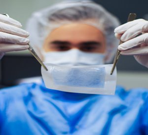 Poietis technician holds up 3D-printed tissue produced in their lab.
Photo: Poietis
The firm’s Next Generation Bioprinting platform features high-resolution, laser-assisted printers with software that controls the 3D organization of cells with cell resolution. It uses automation and robotics to guarantee reproducible tissue manufacturing, giving the printed tissue the functionality required by researchers.
Poietis technician holds up 3D-printed tissue produced in their lab.
Photo: Poietis
The firm’s Next Generation Bioprinting platform features high-resolution, laser-assisted printers with software that controls the 3D organization of cells with cell resolution. It uses automation and robotics to guarantee reproducible tissue manufacturing, giving the printed tissue the functionality required by researchers.
The multimodal platform includes bioextrusion, micro-valve bioprinting along with laser-assisted bioprinting, working with online sensors and machine learning algorithms to ensure that what is designed is printed. For 4D applications, the firm looks to its laser-assisted bioprinting technique. It uses laser pulses sent every nanosecond to deposit microscopic drops of bio ink loaded with cells onto a cartridge. The software controls the ejection as well as droplet volume to accuracies close a picoliter. Poietis claims it can achieve 20 μm resolution at speeds up to 10,000 droplets per second, assisted by a 6-axis robotic arm.
Recommended Reading: VA System Rolls Out 3D Printing
Finally, the company claims it is developing new software to “program tissue self-organization, to anticipate the evolution of bioprinted construct with time,” the step that moves the platform to 4D printing.
While commercial 4D applications and products remain distant, there are flurries of work being done in university labs and research centers. One new technique, developed by researchers in Australia and New Zealand, combines a new 3D printing method with photo-controlled/living polymerization, a chemical process that creates polymers.
The technique, called PET-RAFT for photo-induced electron/energy transfer-reversible addition-fragmentation chain transfer, produces long polymer molecules using visible light, rather than heat. Temperatures above 40 °C kill cells but the new process works in ambient temperatures that may provide greater viability. That could allow the process to be used in tissue engineering or other biomedical applications.
“Controlled polymerization has never been used in 3D and 4D printing before, because the rates of typical controlled polymerization processes are too slow for 3D/4D printing, where the reaction must be fast for practical printing speeds,” said Cyrille Boyer, co-director of the Australian Centre for NanoMedicine, School of Chemical Engineering, at the University of New South Wales Sydney. “After two years of research and hundreds of experiments, we developed a rapid process compatible with 3D printing.
“In contrast to conventional 3D printing, our new method of using visible light allows us to control the architecture of the polymers and tune the mechanical properties of the materials prepared by our process,” he said. “This new process also gives us access to 4D printing and allows the material to be transformed or functionalized, which was not previously possible.”
Boyer’s team demonstrated how functionalization could create a 4D material by modifying a commercial 3D printer and using it to produce a material that changes shape in water. The next step is to print more complex polymers and create objects that respond to different stimuli.
While most 4D projects are still trying to overcome obstacles to commercialization in the lab, some startups have also gotten into the game. In Belgium, Jan Schrooten co-founded Antleron to turn cells into personalized therapies and tissues. Last year, the Leuven-based firm and printer manufacturer 3D Systems began to collaborate on furthering bioprinting solutions for regenerative products.
To do this, Antleron CEO Schooten is establishing the living therapy factory. “By definition it will be a digital factory, it will be a modular complex using a combination of 3D printers, bioreactors, biomaterials, and biologics,” he said. “It is streamlined for a manufacturing workflow that is controlled with a matching quality control system to mitigate risks.”
The goal is to develop new, structured workflows for the company’s core technologies to turn cells in therapies. 3DSystems, which has invested in Antleron, brings printers and a portfolio of 21 biocompatible materials along with its 3D Sprint and 3DXpert software and other equipment to the arrangement.
“There’s a combination of engineering material and original natural material, and we’re looking for ways to make that printable,” said Schrooten. The agreement helps enable Antleron to customize materials and optimize printing strategy.
Bioprinting and regenerative medicine are a newer trend in 3D printing, said Chuck Hull, 3D Systems co-founder and chief technology officer. The firm first stepped into the field with an earlier agreement with United Therapeutics, which is working to build a transplantable lung.
“It poses a lot of cell biologicial questions but also 3D printing questions,” Hull said. “They produce human collagen, and our printing inks are collagen-based. We print scaffolds to be infused with living cells. So it has been a great learning experience. Because of our progress, there are a lot of other tissues we could handle.”
But for 4D applications, it is still early, Schrooten said.
Once researchers print a successful object, they then have to show that they can do it reproducibly. After that, they have to show that they can print at volume and overcome medical and regulatory hurdles. “When this will be a bonafide clinical business is not well-defined,” Hull said.
Developments coming out of university and R&D labs will push the development of 4D printing technology and 4D materials. While that is happening, the next phase will come into greater focus: realizing performance standards and meeting regulatory compliance.
“In a few years the first applications will be there,” said Schrooten. “We will see proof of concept. There’s still a lot of work with standards. There’s a lot of research being done on the millimeter scale, but you need to make it in volume. So getting to commercial will take more time.”
John Kosowatz is senior editor.
SIDEBAR 1
Designer uses 4D flow MRI images to 3D print a functional heart model.
3D printing also has potential as a powerful way to visualize changes over time. In Colorado, for example, technicians and doctors are combining 4D magnetic resonance images of blood flow with 3D printers to produce a multicolored functional heart model.
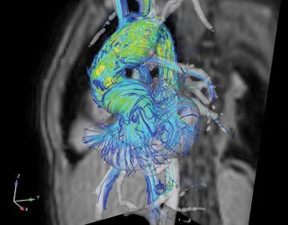 4D flow MRI was sliced to 3D print a heart model showing nuances of blood flow.
Images: Stratasys.
By color coding the velocity at which blood flows through the heart, 4D flow scans give doctors a simple way to visualize exactly where problems are located as blood moves through each section of the heart during a cardiac cycle, from the ending of one heartbeat to the beginning of the next. This helps them pinpoint exactly where problems are located and plan appropriate surgery.
4D flow MRI was sliced to 3D print a heart model showing nuances of blood flow.
Images: Stratasys.
By color coding the velocity at which blood flows through the heart, 4D flow scans give doctors a simple way to visualize exactly where problems are located as blood moves through each section of the heart during a cardiac cycle, from the ending of one heartbeat to the beginning of the next. This helps them pinpoint exactly where problems are located and plan appropriate surgery.
“It is not static, you can actually see the flow come in and out,” said Lorna Browne, a radiologist at Colorado Children’s Hospital. 4D flow scans image every major vessel in the chest cavity. That allows doctors to get a complete read on the patient, something a standard MRI scan does not provide. “It is easier to interpret.”
The printed model is based on a Stratasys digital anatomy solution that includes a suite of new materials the company says more closely mimics actual tissue density rather than a rigid model. “This is more about trying to replicate native tissue,” said Scott Drikakis, medical segment leader for Stratasys. He added that the platform allows for greater and more efficient blending to better approximate varying physical properties of an organ. It becomes a functional model for surgical planning rather than simply a visual model.
Drikakis said the firm worked directly with medical device manufacturers and doctors in developing the platform. That helped in designing tissue as soft and pliable as 00 on the Shore A Hardness scale, the lowest score of a material on the market, he claimed.
To print a functional model, the system relies on voxel-based software and Stratasys’ J750 printer, the first printer the firm designed specifically for a market, according to Drikakis. It works with new software in its GrabCAD package that defines terms on a more clinical level. “It doesn’t talk of Shore values, it talks of bone density,” he said. “Instead of having to figure out what shore values are, you are just talking bone density—more rigid, less rigid—using clinical descriptions.”
Extracting the MRI files for printing was challenging, said Nicholas Jacobson, a clinical surgical design researcher at Colorado Children’s Hospital. “It was quite a process to take that data out of the computer, slice by slice, and send through the printer.”
To prepare the MRI files for printing, Jacobson exported flow lines as colored .PLY files because they store color scalar data in vertices of meshes and export very quickly. Those files were then imported in the modeling software that reads the .PLY files as voxel shape channels and uses color data to color channels of the voxel shapes. “This is important because we can then control the width of the 4D flow lines and more accurately control the gradient of colors,” he said.
Then Jacobson used a dithering algorithm to “pixelize” the colors, dividing them into 15 x 15-micron squares that are the size of a droplet coming out of the printer. “We are literally specifying every drop,” he said.
Dithering, or mixing pixels of several colors together to trick the eye into seeing an entirely different color, was also needed because the printer is limited to seven colors. Algorithms quantify the ratios and patterning of placing materials next to each other.
“If done correctly, it can create a blended look that appears to represent more than 250,000 colors,” Jacobson said.
“Finally, we launch a slicing algorithm that slices the model at .027 mm and exports .PNG files. The printer reads the .PNG files like an inkjet printer. This process is incredibly powerful because of the detail we are getting. But this is still nascent technology. We haven’t done a lot of it yet,” he said.
SIDEBAR 2: Symbiosis in AM Design
Provided by COMSOL
Topology optimization is, as the name suggests, an optimization method that can identify the optimal topology for a given design problem. No initial design is required; instead, the designer inputs a space for the design to exist, a specification of the physics pertaining to the analysis, and a list of constraints and objectives. Based on this information, the optimization algorithm can identify valid designs and quantify their performance in a systematic way.
The method is associated with extreme design freedom and extreme performance, which makes it popular for pushing the boundaries of engineering. Performance aside, the limited input required to come up with new designs also makes the method attractive for generating custom designs with similar specifications, as seen within the biomedical field, where every implant is similar, yet patient specific.
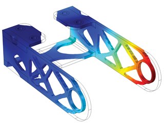 Topology optimization updates the design in an iterative fashion using the gradient of the objective function. Photo: COMSOL
There is a symbiotic relationship between the fields of topology optimization and additive manufacturing. This is due to the fact that topology optimization often produces designs that are too complex for manufacturing with conventional methods. On the other hand, most designers struggle to take advantage of the design freedom that additive manufacturing provides, and thus welcome tools that automatically design for performance. Therefore, the method has become popular within structural mechanics, where the meaning of performance, the objective function, is agreed upon and well-defined.
Topology optimization updates the design in an iterative fashion using the gradient of the objective function. Photo: COMSOL
There is a symbiotic relationship between the fields of topology optimization and additive manufacturing. This is due to the fact that topology optimization often produces designs that are too complex for manufacturing with conventional methods. On the other hand, most designers struggle to take advantage of the design freedom that additive manufacturing provides, and thus welcome tools that automatically design for performance. Therefore, the method has become popular within structural mechanics, where the meaning of performance, the objective function, is agreed upon and well-defined.
Topology optimization updates the design in an iterative fashion using the gradient of the objective function. It is normal to have thousands of design variables, so the gradient has to be computed in an efficient way, and the only way to achieve this is with adjoint sensitivity analysis.
Researchers in academia have demonstrated topology optimization for nonstandard problems outside the field of structural mechanics, so there are no inherent limitations preventing the widespread use of the method.
The derivation of the adjoint sensitivity analysis can, however, be a laborious and difficult task to perform, but the COMSOL Multiphysics® software is capable of performing adjoint sensitivity analysis automatically, which empowers nonexperts to use topology optimization for nonstandard objective functions, regardless of the types of physics. We now see the application of topology optimization within computational fluid dynamics as well as various wave propagation problems, such as acoustics and electromagnetics.
What is the next step? It's 4D printing, a term that is a bit of a misnomer because it still relies on 3D printers. Time is the element that pushes 3D to 4D, creating printed materials that change their shape over time. “You first have to have 3D manufacturing capability and then you add smart materials,” said Howon Lee, an associate professor of mechanical and space engineering at Rutgers University.
Smart materials have the ability to change shape over time, creating a wide-ranging universe of potential new products. NASA has already produced woven metal fabrics that change shape and are foldable. Their potential use ranges from shielding a spacecraft from extreme temperatures to erecting an antenna in space. Airbus is investigating 4D-printed components that could lighten the weight and improve the performance of aircraft.
4D-printed healthcare applications may be the closest to commercial applications. Shape-shifting materials could be used for small, implantable medical devices, said Lee. Tiny, soft devices could be inserted or implanted in people, and harden when they reach the affected area. But with a few possible exceptions, wide-scale application remains years away. Most activity now is still in research and development. The market is in its infancy.
Latest in News: How Coronavirus is Reshaping the Engineering World
One survey by ResearchandMarkets.com puts the overall market for 4D printing in healthcare at a meager $9 million in 2021, rising to $32 million by 2026. In 2021, the medical and research models segment is expected to account for the largest share of the overall healthcare market. “The ability of the 4D printing technology in manufacturing smart medical models will bring significant transformation in the medical field and will support the growth of this segment in the forecast period,” according to the report. Yet, despite its small market size, emerging research shows that 4D printing could provide physicians and surgeons with new tools for everything from generating replacement skin to creating objects that respond to changes in their environment.
First Results
One example is work taking place at the Self-Assembly Lab at the Massachusetts Institute of Technology. Using Autodesk’s Cyborg software and Stratasys 3D printers, it has been developing materials for more than five years that change shape when triggered by water.
MIT's Self-Assembly Lab employs technology that prints "smart" self-folding materials that can transform shape. Photo: Courtesy of Self-Assembly Lab, MIT + Stratasys Ltd + Autodesk Inc.
Skylar Tibbets runs MIT’s Self-Assembly Lab, and may be the first to delve into what is now called 4D printing. In 2013, he declared an “unprecedented revolution” taking place at the micro and nano level to program physical and biological materials to change shape and form. The lab is working toward self-forming structures to eventually impact infrastructure and construction. “It just might be the manufacturing technique that allows us to produce more adaptive infrastructure in the future,” he said.
Perhaps the earliest successful applications will come in the areas of tissue regeneration or regenerative medicine. 3D Systems, Stratasys, and other printer manufacturers see the segment growing over the next few years and are working with biotech and bioprinting firms eager to adapt the technology to processes and production.
Bioprinting is a process of printing materials with living cells, most often for product testing and, further out, reconstructive surgery. Because the printed objects contain living cells, they grow and morph into living tissue over time. Poietis, a French bioprinting firm developing its own 4D printing platform, was the first to commercialize a bioprinted synthetic skin, Poieskin.
Poietis is still a long way from providing skin tissue for reconstructive surgery, but its 3D-printed skin has found a place in cosmetic testing, exploiting the movement condemning and in some cases outlawing the use of animals for testing. In 2018, it signed an agreement with Servier, a pharmaceutical company, to develop a 4D-bioprinted liver model to better predict drug toxicity.
Poietis technician holds up 3D-printed tissue produced in their lab.
Photo: Poietis
The multimodal platform includes bioextrusion, micro-valve bioprinting along with laser-assisted bioprinting, working with online sensors and machine learning algorithms to ensure that what is designed is printed. For 4D applications, the firm looks to its laser-assisted bioprinting technique. It uses laser pulses sent every nanosecond to deposit microscopic drops of bio ink loaded with cells onto a cartridge. The software controls the ejection as well as droplet volume to accuracies close a picoliter. Poietis claims it can achieve 20 μm resolution at speeds up to 10,000 droplets per second, assisted by a 6-axis robotic arm.
Recommended Reading: VA System Rolls Out 3D Printing
Finally, the company claims it is developing new software to “program tissue self-organization, to anticipate the evolution of bioprinted construct with time,” the step that moves the platform to 4D printing.
Back in the Lab
While commercial 4D applications and products remain distant, there are flurries of work being done in university labs and research centers. One new technique, developed by researchers in Australia and New Zealand, combines a new 3D printing method with photo-controlled/living polymerization, a chemical process that creates polymers.
The technique, called PET-RAFT for photo-induced electron/energy transfer-reversible addition-fragmentation chain transfer, produces long polymer molecules using visible light, rather than heat. Temperatures above 40 °C kill cells but the new process works in ambient temperatures that may provide greater viability. That could allow the process to be used in tissue engineering or other biomedical applications.
“Controlled polymerization has never been used in 3D and 4D printing before, because the rates of typical controlled polymerization processes are too slow for 3D/4D printing, where the reaction must be fast for practical printing speeds,” said Cyrille Boyer, co-director of the Australian Centre for NanoMedicine, School of Chemical Engineering, at the University of New South Wales Sydney. “After two years of research and hundreds of experiments, we developed a rapid process compatible with 3D printing.
“In contrast to conventional 3D printing, our new method of using visible light allows us to control the architecture of the polymers and tune the mechanical properties of the materials prepared by our process,” he said. “This new process also gives us access to 4D printing and allows the material to be transformed or functionalized, which was not previously possible.”
Boyer’s team demonstrated how functionalization could create a 4D material by modifying a commercial 3D printer and using it to produce a material that changes shape in water. The next step is to print more complex polymers and create objects that respond to different stimuli.
Commercialization
While most 4D projects are still trying to overcome obstacles to commercialization in the lab, some startups have also gotten into the game. In Belgium, Jan Schrooten co-founded Antleron to turn cells into personalized therapies and tissues. Last year, the Leuven-based firm and printer manufacturer 3D Systems began to collaborate on furthering bioprinting solutions for regenerative products.
To do this, Antleron CEO Schooten is establishing the living therapy factory. “By definition it will be a digital factory, it will be a modular complex using a combination of 3D printers, bioreactors, biomaterials, and biologics,” he said. “It is streamlined for a manufacturing workflow that is controlled with a matching quality control system to mitigate risks.”
The goal is to develop new, structured workflows for the company’s core technologies to turn cells in therapies. 3DSystems, which has invested in Antleron, brings printers and a portfolio of 21 biocompatible materials along with its 3D Sprint and 3DXpert software and other equipment to the arrangement.
“There’s a combination of engineering material and original natural material, and we’re looking for ways to make that printable,” said Schrooten. The agreement helps enable Antleron to customize materials and optimize printing strategy.
Bioprinting and regenerative medicine are a newer trend in 3D printing, said Chuck Hull, 3D Systems co-founder and chief technology officer. The firm first stepped into the field with an earlier agreement with United Therapeutics, which is working to build a transplantable lung.
“It poses a lot of cell biologicial questions but also 3D printing questions,” Hull said. “They produce human collagen, and our printing inks are collagen-based. We print scaffolds to be infused with living cells. So it has been a great learning experience. Because of our progress, there are a lot of other tissues we could handle.”
But for 4D applications, it is still early, Schrooten said.
Once researchers print a successful object, they then have to show that they can do it reproducibly. After that, they have to show that they can print at volume and overcome medical and regulatory hurdles. “When this will be a bonafide clinical business is not well-defined,” Hull said.
Developments coming out of university and R&D labs will push the development of 4D printing technology and 4D materials. While that is happening, the next phase will come into greater focus: realizing performance standards and meeting regulatory compliance.
“In a few years the first applications will be there,” said Schrooten. “We will see proof of concept. There’s still a lot of work with standards. There’s a lot of research being done on the millimeter scale, but you need to make it in volume. So getting to commercial will take more time.”
John Kosowatz is senior editor.
SIDEBAR 1
Designer uses 4D flow MRI images to 3D print a functional heart model.
3D printing also has potential as a powerful way to visualize changes over time. In Colorado, for example, technicians and doctors are combining 4D magnetic resonance images of blood flow with 3D printers to produce a multicolored functional heart model.
4D flow MRI was sliced to 3D print a heart model showing nuances of blood flow.
Images: Stratasys.
“It is not static, you can actually see the flow come in and out,” said Lorna Browne, a radiologist at Colorado Children’s Hospital. 4D flow scans image every major vessel in the chest cavity. That allows doctors to get a complete read on the patient, something a standard MRI scan does not provide. “It is easier to interpret.”
The printed model is based on a Stratasys digital anatomy solution that includes a suite of new materials the company says more closely mimics actual tissue density rather than a rigid model. “This is more about trying to replicate native tissue,” said Scott Drikakis, medical segment leader for Stratasys. He added that the platform allows for greater and more efficient blending to better approximate varying physical properties of an organ. It becomes a functional model for surgical planning rather than simply a visual model.
Drikakis said the firm worked directly with medical device manufacturers and doctors in developing the platform. That helped in designing tissue as soft and pliable as 00 on the Shore A Hardness scale, the lowest score of a material on the market, he claimed.
To print a functional model, the system relies on voxel-based software and Stratasys’ J750 printer, the first printer the firm designed specifically for a market, according to Drikakis. It works with new software in its GrabCAD package that defines terms on a more clinical level. “It doesn’t talk of Shore values, it talks of bone density,” he said. “Instead of having to figure out what shore values are, you are just talking bone density—more rigid, less rigid—using clinical descriptions.”
Extracting the MRI files for printing was challenging, said Nicholas Jacobson, a clinical surgical design researcher at Colorado Children’s Hospital. “It was quite a process to take that data out of the computer, slice by slice, and send through the printer.”
To prepare the MRI files for printing, Jacobson exported flow lines as colored .PLY files because they store color scalar data in vertices of meshes and export very quickly. Those files were then imported in the modeling software that reads the .PLY files as voxel shape channels and uses color data to color channels of the voxel shapes. “This is important because we can then control the width of the 4D flow lines and more accurately control the gradient of colors,” he said.
Then Jacobson used a dithering algorithm to “pixelize” the colors, dividing them into 15 x 15-micron squares that are the size of a droplet coming out of the printer. “We are literally specifying every drop,” he said.
Dithering, or mixing pixels of several colors together to trick the eye into seeing an entirely different color, was also needed because the printer is limited to seven colors. Algorithms quantify the ratios and patterning of placing materials next to each other.
“If done correctly, it can create a blended look that appears to represent more than 250,000 colors,” Jacobson said.
“Finally, we launch a slicing algorithm that slices the model at .027 mm and exports .PNG files. The printer reads the .PNG files like an inkjet printer. This process is incredibly powerful because of the detail we are getting. But this is still nascent technology. We haven’t done a lot of it yet,” he said.
SIDEBAR 2: Symbiosis in AM Design
Provided by COMSOL
Topology optimization is, as the name suggests, an optimization method that can identify the optimal topology for a given design problem. No initial design is required; instead, the designer inputs a space for the design to exist, a specification of the physics pertaining to the analysis, and a list of constraints and objectives. Based on this information, the optimization algorithm can identify valid designs and quantify their performance in a systematic way.
The method is associated with extreme design freedom and extreme performance, which makes it popular for pushing the boundaries of engineering. Performance aside, the limited input required to come up with new designs also makes the method attractive for generating custom designs with similar specifications, as seen within the biomedical field, where every implant is similar, yet patient specific.
Topology optimization updates the design in an iterative fashion using the gradient of the objective function. Photo: COMSOL
Topology optimization updates the design in an iterative fashion using the gradient of the objective function. It is normal to have thousands of design variables, so the gradient has to be computed in an efficient way, and the only way to achieve this is with adjoint sensitivity analysis.
Researchers in academia have demonstrated topology optimization for nonstandard problems outside the field of structural mechanics, so there are no inherent limitations preventing the widespread use of the method.
The derivation of the adjoint sensitivity analysis can, however, be a laborious and difficult task to perform, but the COMSOL Multiphysics® software is capable of performing adjoint sensitivity analysis automatically, which empowers nonexperts to use topology optimization for nonstandard objective functions, regardless of the types of physics. We now see the application of topology optimization within computational fluid dynamics as well as various wave propagation problems, such as acoustics and electromagnetics.
Related Content
Breakthrough Could Make for Long-Range EVs
Apr 16, 2024
A simple method could lead to electric vehicles that can go much father on a single charge, and batteries that last years longer than present technology.
BatMan Breakthrough in EV Battery Manufacturing
Mar 28, 2024
Project looks to optimize battery charging speeds in EVs using laser ablation, advanced computational models, and materials characterization.

New 3D Printing Method Cuts Material Development Time in Half
Feb 12, 2024
3D Mesh Wearables Transmit Data, No Wi-Fi Required
Feb 6, 2024
A new set of wearables will make digital health more accessible by not requiring Wi-Fi or cellular connectivity to relay information over long distances.